A Review of the Nature and Origin of Limestone Microporosity
1. Introduction
Carbonates and sandstones form important reservoirs for hydrocarbons and can act as potential sinks for the future storage of anthropogenic green-business firm gases by carbon capture and sequestration. The nature of the pore infinite in these stone types tin can be circuitous, often resulting from a combination of depositional and diagenetic processes. The pore geometry of carbonates is ordinarily more than tortuous due to millimeter calibration macroporosity, which may include interparticle, intercrystal, moldic and vuggy porosity (Choquette and Pray, 1970), and microporosity (submicron porosity), typically intraparticle, including partially dissolved bioclastic and/or grain material, and cementation found along the pore and throat grain contacts. Clean sandstones display a simplistic pore geometry, with macroporosity forming a more than uniform intergranular network and microporosity forming from detrital and authigenic clays (Pittman, 1979). "Tight" (low porosity) sandstones, however, can demonstrate characteristics that lead to a more than complicated pore network. Such features include variable grain size, grain packing, sorting, clay distribution and after burial compaction (Marquez et al., 2014; Teles et al., 2016). Additionally, main pore geometry in these sandstones can exist reduced past cementation, rendering them "tight." Consequently, determination of petrophysical properties of these sandstones are oft as challenging as carbonate rocks.
Conventional relationships used to predict petrophysical properties are restricted past the presence of microporosity found in many carbonates and tight sandstones. Such relationships include Archie'due south law for resistivity, the Carman–Kozeny permeability guess and the Brooks–Corey parameterization of relative permeability, which were all originally designed for more simplistic rocks with intergranular pores. When considering these classic models for complex carbonates and tight sandstones, the results are inaccurate due to the greater range in pore-size distributions and interconnectivity of different pore types (Jennings and Jerry Lucia, 2003).
The use of X-ray computed microtomography (micro CT), high resolution, 3-dimensional (3D) images of porous geological media accept become profoundly pop over the past 2 decades (Valvatne and Edgeless, 2004; Andrä et al., 2013a,b; Madonna et al., 2013; Prodanović et al., 2014; Bultreys et al., 2016a). This not-invasive technique allows 3D pore geometries to be evaluated quantitatively, whilst preserving the integrity of the original sample. The other great advantage of a digitized pore space network reconstruction is the ability to simulate a multitude of different ship properties. Porous flow, in detail, has several applications to many important geological fields, including hydrology, the recovery of oil and gas, and carbon sequestration. Recent studies related to the storage of anthropogenic CO2 in the subsurface highlight the in situ process of dissolution and its effects on porosity and permeability of different carbonates by means of experiment performed with lab-based micro CT scanner visualization (Menke et al., 2015, 2016). Numerical models of single and multiphase fluid flow take also been used to predict accented and relative permeability for a diversity of dissimilar rock type. The employ of traditional computation fluid dynamics, which incorporates finite difference, finite book and finite element (Mostaghimi et al., 2013; Raeini et al., 2014), and the lattice-Boltzmann method (Ramstad et al., 2010; Shah et al., 2016) have proven popular in recent years. For more particular on the use of micro CT and its applications to geoscience, the reader is referred to a number of recent review papers (Cnudde and Boone, 2013; Bultreys et al., 2016a; Berg et al., 2017).
Other studies have shown that micro CT can provide useful insights into the central processes occurring at the pore scale inside unconventional resources such as shale oil and shale gas. Ma et al. (2016) successfully combined micro CT and scanning electron microscopy (SEM) to identify and quantify four dissimilar types of porosity inside the Bowland Shale, United kingdom. Their results indicated that porosity was unconnected at scales greater than xx nm, whilst organic matter and clay minerals at scales of less than 20 nm was found to exist connected and offered potential diffusion transport pathways for gas. A similar study by Saif et al. (2017) on the Greenish River oil shale (USA) highlighted the evolution of an interconnected fracture network and increased porosity due to the breakdown of kerogen into hydrocarbon fluids during pyrolysis. Such insights are powerful tools which can be used to judge the petrophysical properties of many rocks for modeling and designing production processes.
Microporosity typically exists at a resolution beneath that from which clear partitioning tin can non be accomplished. This results in an intermediate phase, that can neither be assigned to void space nor solid in conventional micro CT. To accurately visualize and represent microporosity in 3D it is possible to combine SEM with various reconstruction techniques (Øren and Bakke, 2003; Okabe and Edgeless, 2005), or more than recently, with the use of focused ion axle scanning electron microscopy (FIB-SEM) (Bera et al., 2011; Jiao et al., 2014). The variation in pore sizes between micro and macroporosity means that non all pores can be incorporated in one unmarried imaging experiment. It remains challenging to combine data from multiple experiments because the necessary resolution of such a model is influenced by the finest pores, while the minimal size of the model is controlled by the representative elementary volume of the sample (Bultreys et al., 2016a). The joint modeling of such different scales results in a difference of several orders of magnitude, which ofttimes presents computational limitations. The use of a pore network model (PNM) reduces the computational expense, which at present are probably the best suited technique for dual calibration porosity simulations (Jiang et al., 2012, 2013; Mehmani and Prodanović, 2014; Bultreys et al., 2015).
Despite the challenges in identifying and quantifying microporosity, their presence can provide an insight into the nature of fluid flow through a continued, intergranular macroporous network. In this article, nosotros distinguish microporosity arising from 2 different sources: microporosity in primary stone-forming grains and microporosity within secondary phases. An example of the former includes fossils in carbonate rocks while an example of the latter includes microporous cement deposited equally secondary phases forth the walls of a pore network. Recent work by Bultreys et al. (2015) discussed the constructive transport properties combining imaged macroporosity and models of microporosity. We refer the reader to this work for physical properties involving microporosity and focus on (a) the pore network geometry of the connected macro pore space, macroporosity, and (b) the manner evolution of secondary microporosity, or cement, impact the connectivity of the primary porosity.
We segment and clarify the micro CT images of half dozen natural sandstones and carbonates into three phases (microporosity, macroporosity and matrix) to quantify their book fraction and to evaluate the nature of their pores and throats using pore network models. We further analyze each rock type by selecting ane or more than smaller regions of interest (ROI) to assess sample heterogeneity and perform fault analysis. We compare the results between three dissimilar sandstones, the Doddington, Knorringfjellet, and Wilcox, and 3 different carbonates, the Estaillades, Massangis Jaune, and Savonnières. The characteristics of the raw data for all vi samples is summarized in Table 1.
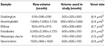
Tabular array 1. Characteristics of raw data.
2. Methods
All data used in this work was sourced externally. The Doddington and Wilcox sandstone data sets were acquired online from the British Geological Club's (BGS) National Geoscience Information Centre (Shah et al., 2016) and the Digital Rocks Portal (Mehmani and Prodanović, 2014; Prodanović et al., 2014), respectively. Images for the Knorringfjellet sandstone and three carbonate samples, Estaillades, Massangis Jaune (Boone et al., 2014), and Savonnières were kindly provided by Tom Bultreys (Bultreys et al., 2016b) (Majestic Higher London).
We filtered the gray scale images to reduce noise and remove unwanted artifacts. We then segmented the filtered prototype into microporosity, macroporosity and matrix by thresholding. Side by side, we analyzed the proportion of microporosity and macroporosity in each sample, using a series of ROI to assess the influence of sample heterogeneity. Pore network modeling and absolute permeability simulations were then carried out to characterize the nature of the pore geometries in all samples and to quantify the outcome of microporosity on single phase fluid menstruum. We used the commercial software PerGeos, from Thermo Fisher Scientific, to prepare images, to perform network modeling, and to simulate fluid flow through the segmented 3D images.
two.1. Image Processing
We filtered the micro CT images to reduce spurious features that occur from the epitome conquering procedure (Ketcham and Carlson, 2001; Wildenschild et al., 2002) and to dilate the contrast between primal phases. Firstly, we extracted a subvolume from the original raw data for computational efficiency in filtering and simulations at a later stage. The sub volume characteristics are highlighted in Table 1. Next, nosotros applied various filters including anisotropic diffusion, non-local means, and despeckle to reduce salt and pepper noise at the eye of grains, whilst maintaining detail at the boundaries between pores and solid matrix.
The process of partitioning in micro CT images is a crucial footstep which tin induce operator bias and later impact the quality of all other image analyses (Wildenschild and Sheppard, 2013; Schlüter et al., 2014). A wide variety of division techniques exist, which can broadly exist separated into two groups. Global thresholding includes all methods where labels are assigned to voxels past histogram evaluation just, without idea for how the gray values are spatially arranged in the paradigm. Such techniques have been reviewed extensively in the past by many authors (Pal and Pal, 1993; Trier and Jain, 1995; Sezgin and Sankur, 2004; Iassonov et al., 2009), only to conclude that none of the methods are particularly good for all segmentation challenges. Local segmentation techniques make up the other popular class and these take account of neighborhood statistics enabling smoother boundaries, avoidance of noise objects, and compensation for local intensity changes. This greater flexibility and often more satisfying segmentation results is what differentiates the local segmentation methods from global thresholding (Iassonov et al., 2009; Wang et al., 2011).
In this piece of work we used manual thresholding to segment the book into three phases: microporosity, macroporosity and matrix. Whilst the classification for labeling the macroporosity and matrix were adequately unproblematic, the microporosity existed every bit an intermediate phase, which created a more challenging interpretation. This intermediate phase included voxels of a particular grayness scale that could often be identified in contact with macroporosity at the edges of grains (cementation), and sometimes occurring as whole or part of grains. This observation suggests that some microporosity exists as partially dissolved grains and/or bioclastic fabric in nearly of the samples. Although our chosen technique was user dependent and was likely to result in a certain degree of incertitude, we manually adjusted the thresholds to produce results that we considered visually correct. To further reduce this uncertainty we benchmarked our results where possible with those found in previous studies (Boone et al., 2014; Derluyn et al., 2014; Mehmani and Prodanović, 2014; Alyafei and Blunt, 2016; Bultreys et al., 2016b). The micrographs in Figure 1 show the segmented gray scale images from the six samples analyzed in this study.
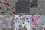
Figure 1. From acme, left to correct: (a) Doddington sandstone, (b) Knorringfjellet sandstone, (c) Wilcox sandstone, (d) Estaillades limestone, (e) Massangis Jaune limestone, and (f) Savonnières limestone. Single slice view of each sample. A quarter of the image is represented in gray scale only. The remaining voxels display macroporosity in ruby, and microporosity in blueish.
two.2. Pore Network Model (PNM)
Following segmentation, pore-based PNM are extracted using a hybrid method described in Youssef et al. (2007). This method employs a medial-axis algorithm guided by a distance map which progressively erodes the pore-grain interface until a one-voxel thick skeleton of the unabridged pore infinite is accomplished. The algorithm then calculates the length and connectivity of each line and designates each one equally either pore or throat based on a known extreme radius. The intersection between two or more lines is marked as a node. A segment of the skeleton is marked equally a pore if the extreme radius of the segment is larger than its length, whilst throats are classified as segments with length that do not exceed their extreme radius. The subsequent expansion of the one-voxel thick skeleton in these pores and throats allows the calculation of the radius of sphere (able to fit in each pore) and the length and equivalent hydraulic radius of each throat. The output of this algorithm results in the coordination number (number of throats connected to each pore), radius, area and volume of every pore, and the radius and length of each throat.
We test a simple model to evaluate the effects of microporosity on the pore geometry using two different scenarios. Initially, we generate the PNM using only the connected pore infinite (where possible) of macroporosity. Post-obit this, we generate a 2d PNM where we presume that microporosity is made up of 100% void infinite, thus describing the pore network prior to cementation. To do this, nosotros simply created a separate binary paradigm for the total porosity case which incorporated both the microporosity and macroporosity. In samples where microporosity occurs within the cement, this step allows us to compare the reduction of pore connectivity and permeability due to cementation. All PNM simulations reported here are derived only from the macropores. The PNM analysis of micropores is an interesting topic for research involving high resolution nano-CT.
2.2.1. Porosity
Using the smaller sub-sampled volumes of each rock, nosotros generated new characterization fields for microporosity and macroporosity in each volume. Microporosity was assigned to the voxels deemed to be associated with the primary rock-forming fabric (east.g., fossils in carbonates that may have been affected by secondary dissolution) and cement phases institute along the walls of the pores. Typically these phases appear as a lighter shade of grey compared to macroporosity. The macroporosity was assigned with much less difficulty based on a dark black or gray color (Figure 1). The volume fraction of porosity was calculated in each of these by measuring the number of voxels assigned to the void space, compared to those making up the remaining groundwork voxels in each sub-sample. The macroporosity and microporosity proportion was calculated for each of the different samples by using the phase segmented data and computing the volume of the total number of macroporosity and microporosity voxels to the total number of voxels in the complete sample. The total porosity was simply calculated by calculation the macroporosity and microporosity. The volumes used for the calculations of the entire sample are highlighted in Table 1, whilst the volume fraction of porosity is summarized in Table 2.

Table 2. Book fraction of total porosity (ϕ T ), macroporosity (ϕ M ), microporosity (ϕ m ), total connected porosity (ϕ T C ), connected macroporosity (ϕ M C ), and connected microporosity (ϕ grand C ) of total smple volumes.
We took the porosity analysis a step farther and separated each sample down into further divisions to appraise the heterogeneity at this scale. All samples were subsampled forth the Z-axis into divisions of three, with the exception of the Knorringfjellet sandstone (8 subvolumes). Effigy two shows a volume rendering representation of each sample and their subdivisions. The data from this analysis is summarized in the Supplementary Material.
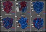
Figure 2. Book rendering visualization of the six different samples. From height, left to right: (a) Doddington sandstone showing connected macroporosity and total microporosity, (b) Knorringfjellet sandstone showing connected macroporosity and connected microporosity, (c) Wilcox sandstone showing total macroporosity and total microporosity, (d) Estaillades limestone showing continued macroporosity and connected microporosity, (eastward) Massangis Jaune limestone showing total macroporosity and continued microporosity, and (f) Savonnières limestone showing continued macroporosity and connected microporosity. For each volume the macroporosity is shown in cherry and microporosity in blue. Table 2 summarizes the volume fractions of porosity observed in all samples. Subdivisions are besides shown here, from which the subvolume porosity analysis was performed.
It is important to note that microfractures tin contribute toward the connectivity of pores in many rocks. These microfractures may exist natural or induced during the sample preparation stage. With the images used in this study we found no prove to suggest the presence of microfractures, and so focus on the primary porosity that can exist observed in all images. The connectivity of the macroporosity in each subsample volume was assessed along the Z-axis. Volumes plant to accept a connected network that passes a percolation threshold from top to lesser were classified as having a connected porosity. Voxels that vest to the initial porosity only exercise not make up part of the continued network are adamant as isolated pores and are removed from the resulting binary image. To fully evaluate the heterogeneity of each stone we generated a combined (microporosity and macroporosity) label field to test the full porosity case and assess whether or non the microporosity had any upshot on the connectivity of each sub-sample volume.
2.2.two. Permeability Simulation
Nosotros performed absolute permeability simulations on each rock to summate the flow of fluid directly through the continued pore space of the 3D micro CT images. We used a finite book solver in the petrophysics module of PerGeos to summate the flow of water given by Stokes equation,
where u is the fluid velocity vector, P is the force per unit area and μ = 1 × x−three Pa.s is viscosity of water. Each simulation was set upwardly with a series of specified boundary conditions described in Thomson et al. (2018). Parameters were kept constant for all simulations, with the exception of the 3D micro CT input prototype.
Nosotros carried out numerical simulations on the connected pore network derived from both the total pore space and the macro pore space for all samples except for the Wilcox sandstone and Massangis Jaune carbonate. In the former, a connected pore network could non be established using the method described in a higher place. In the latter, a continued network was absent-minded in macropores, but existed in micropore and total pore space. Samples in which micropores occur within the cement phase, we define the normalized permeability , where k M is the permeability derived from the macro pore network and 1000 T is the permeability derived from the total pore infinite (eastward.grand., both macro and micro pores combined together). In section iv we show the variations in g* with cement book fraction (volume fraction of micropores).
Nosotros commencement carried out permeability simulations in samples with a continued porosity beyond the unabridged volume, evaluating both one thousand M and thousand T , to assess the changes to permeability with the add-on of microporosity permitting total flow. Side by side, nosotros followed a similar approach to appraise the influence of heterogeneity within the microtomographic images. Nosotros selected minor regions of involvement in each volume forth their Z-axis. Each subvolume was 200 slices (voxels) in length. We then calculated the total porosity and connected porosity, when possible, in each sub-sample. Volumes found to have connected porosity were used to simulate permeability. Variations in porosity within the small subvolumes are shown in Figures 3B,D for Doddington and Knoringfjellet sandstones.
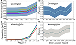
Figure three. (A) Plot of calculated permeability as a function of the balance from permeability simulations in Doddington Sandstone. (B) Variation of porosity measured within subvolumes of a sample of Doddington. (C,D) Same as in panels (A,B) respectively, from Knorringfjellet. The shaded region indicates the maximum and minimum values obtained from each subvolume.
The numerical value of permeability, determined from the simulations, depends strongly on the convergence criterion for the simulation. The convergence criterion or error, ϵ, is defined equally the maximum change in the value of an unknown variable (e.g., velocity and pressure) per time pace. Equations (one) and (2) describe a steady-state, i.e., the calculates values of x, y, and z components of velocity and pressure should non change on subsequent iterations. As a result, we tin define,
where n is the current iteration, v i are components of the velocity vector, P is the pressure, t is the time pace and c two is a compressibility coefficient used in the simulation.
We carried out a series of numerical experiments (Figures 3A,C), by terminating the simulations when a prescribed value of ϵ is reached. The results in Figures 3A,C) show the sensitivity of the permeability to the value of ϵ. While a loftier value of ϵ, such every bit ten−three, reduces the computation fourth dimension, the computed permeability is significantly higher than those obtained for a lower value of ϵ. For example, in Figures 3A,C, the calculated permeability remained nearly abiding for ϵ ≤ 10−5. After performing this initial series of sensitivity tests on Doddington and Knoringfjellet sandstones, we prepare the value of ϵ between ten−5 and 10−half dozen for the remaining permeability simulations.
iii. Results
For each sample we compare the porosity, PNM characteristics and permeability. We testify the total porosity, the proportion of macroporosity and microporosity, and the volume of connected (effective) porosity for each of these. The PNM attributes highlight the variation in the characteristics of pores and throats. Lastly, we compare the results of permeability simulations using a macroporosity merely model and a total porosity (micro and macro) model through the samples.
3.1. Porosity Analysis
The Doddington and Knorringfjellet sandstones are both potential COtwo sequestration target reservoirs with quite different porosity characteristics. The Doddington sandstone has a higher full porosity of 18.6%, which is largely shared by macroporosity, contributing to 89.8% of the pore space, whilst but a small fraction of microporosity makes up the remaining network. Overall, the Doddington sandstone has good effective porosity with total connected porosity at eighteen.5% and connected macroporosity at 16.half dozen%. Conversely, the Knorringfjellet sandstone has a much lower total porosity of 7.seven%. The macroporosity and microporosity in this sample are 4.three and 3.iv%, respectively, indicating that the microporosity occupies a larger proportion (44.ii%) of the total pore network compared to that observed in the Doddington sandstone (10.2%). The total connected porosity observed in the Knorringfjellet sandstone is 20% reduced compared to its overall total porosity, significantly more compared to that seen in the Doddington sandstone.
The unconventional Wilcox sandstone is a tight gas target with similar porosity characteristics to the Knorringfjellet sandstone. The Wilcox also has a low full porosity of only 7.1%, separated by 3.ii% macroporosity and 3.9% microporosity. In this sample the microporosity contributes toward 54.ix% of the full pore space, providing the highest proportion of microporosity observed in all the sandstone samples that we analyzed. As indicated in the information in Tabular array 2, we failed to detect an interconnected pore network in this sample.
The Doddington sandstone shows very minimal variation across the three volumes with a range in total porosity from 18 to 19.ii%. The total porosity is reduced by an boilerplate of 0.7% when assessing the connectivity of the full pore volume. The Knorringfjellet sandstone however, shows greater variation across the 8 volumes that were analyzed. This sample has a range in total porosity from 7 to viii.8%, whilst the proportion of connected total porosity is reduced by 20% on boilerplate. The Wilcox sandstone follows a similar trend with increasing heterogeneity. This sample has a total porosity range from 6.6 to 7.4%, and a significant reduction in the proportion of the total porosity when considering the connectivity of the network. On average, the total connected porosity is reduced by 66%. Interestingly, this is only observed in the sub-volume analysis, whilst nada connected porosity was identified using the whole book.
The 3 limestone samples are from outcrop in France. The Estaillades limestone has a full porosity of 19.1%, which is largely occupied by macroporosity, 61.three% of the total pore volume. The microporosity makes up only 7.4% of the total sample book, the lowest proportion observed in all three of the carbonates analyzed. The connectivity of the full porosity is high, with only a 3.7% reduction compared to the total pore volume. The Massangis Jaune carbonate has the lowest total porosity of all three carbonates, with 16.1% total pore volume. Of this, the macroporosity makes upwards only 30.4%, the everyman proportion observed in the carbonates. This sample is also the but one to accept a microporosity fraction that exceeds that of the macroporosity. The total porosity is well-connected, with microporosity seemingly contributing almost entirely toward this. The complete absence of connectivity in macroporosity supports this observation. The Savonnières carbonate is composed largely of ooliths and some consummate and partial shell fragments. This composition provides the highest total porosity observed in all of the samples (32.5%). The macroporosity has a 64.3% share of the full pore book, which appears equally intergranular and moldic porosity. Microporosity fulfills the remaining pore space equally partially dissolved ooliths and trounce material. The full porosity is well-connected, represented past 28.8% of total sample book.
The Estaillades limestone shows the widest range in total porosity, with values from 18.6 to 22.7%. This porosity is reduced by an boilerplate of iii.ix% when we take the connectivity of each pore volume. This is the lowest reduction in porosity observed between the three carbonate samples. The Massangis Jaune sample had a range in total porosity from 15.2 to 17.2% across the sub-volumes. The total continued porosity was ix.7% less on average compared to the total pore book when analyzed. This sample has a huge variation in the connectivity of the macroporosity, with a range from 47.8 to 93.8%. This is interesting as zero connected macroporosity is observed when considering the whole sample volume. The Savonnières limestone has a total porosity range from 31.7 to 33.two%. The connected proportion of this porosity is reduced by 12.half dozen% on average.
3.2. Pore Network Model Characteristics
The PNM uses the macroporosity of each sample to generate an idealized network of pores and throats, represented by red spheres and gray cylinders, respectively (Figure 4). The visualizations shown in Figures 4a–f highlight the dissimilarity between the nature of the pore networks when the models have been scaled to show the same characteristics (pore and throat radius).
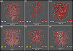
Figure 4. Pore network model visualization of the six samples using the macroporosity network just. From peak, left to right: (a) Doddington sandstone, (b) Knorringfjellet sandstone, (c) Wilcox sandstone, (d) Estaillades limestone, (east) Massangis Jaune limestone, and (f) Savonnières limestone. In this schematic the pores are displayed as red spheres and the throats are greyness cylinders. CN = hateful coordination number, whilst the scarlet text represents the median pore radius and the greyness text represents the median throat radius.
We compare the characteristics of the macropore geometry for the three different sandstone samples: Doddington, Knorringfjellet, and Wilcox in Figures 4a–c. The pore network of Doddington sandstone (Figure 4a) is dense and well-connected, due to higher porosity and by and large larger pores and throats. The mean coordination number for pores is 4, whilst the median pore radius, throat radius and throat length is 25.two, fourteen.nine, and 158.6 μm, respectively. The pore network shown in the Knorringfjellet sandstone (Figure 4b) is significantly less dense, and appears to have a more isolated confined nature compared to the other 2 sandstones. The hateful coordination number is 3, whilst the dimensions of the network in the Knorringfjellet sandstone are the smallest observed in all iii sandstones, with median pore radius of 6.2 μm, throats of radius 3.6 μm and throat length of 33.8 μm. The Wilcox sandstone (Figure 4c) has the lowest connectivity with a mean pore coordination number of one. The depression connectivity of the pore network in Wilcox tin can be visualized in Figure 4c by the relatively loftier affluence of red, spherical pores and depression abundance of gray, cylindrical throats compared to the Doddington network in Figure 4a, which shows a high abundance of throats connecting the pores. In Wilcox, pores have a median radius of vii.4 μm, whilst the median throat radius is 5.v μm and the median pharynx length is 37.i μm.
The macropore network characteristics of the iii different carbonates: Estaillades, Massangis Jaune, and Savonnières are compared in Figures 4d–f. The Estaillades limestone (Figure 4d) has the highest density of small, grayness, and cylindrical throats, reflected by a pore coordination number of v, the highest value observed in all samples. The median pore radius, throat radius and throat length is 12.4, 5.9, and 40.4 μm, respectively. The pore geometry observed in the Massangis Jaune limestone (Figure 4e) is the least well-connected, highlighted by a mean pore coordination number of 1. This sample has median pore radius of 7.two μm, median pharynx radius of 7.9 μm, and median throat length of 53.four μm. The Savonnières limestone network (Effigy 4f) also consists of high throat density, reflected by a pore coordination number of 4. The pores in Savonnières have a median radius of nineteen.2 μm, whilst the throats have a median radius of 9.8 μm, the highest values observed in all three carbonates. The same can be said for the median throat length, calculated at 66.7 μm.
The log normal plots in Effigy v show the distributions of pore and throat geometries in sandstones (Figures 5A,B) and carbonates (Figures 5C,D). Nosotros as well report the mean coordination number, median pore and throat radius, and median throat length values in Table 3. In Figures 5A,B, we included published information from Fontainebleau and Berea Sandstones (Thomson et al., 2018) for comparison. The high coordination number and connectivity in the Doddington sandstone is conspicuously demonstrated by the peaks in the histograms in Figures 5A,B. The distribution of pore radius in all three carbonates in Figure 5C are asymmetric with a skewness toward the right. In comparison, distribution of throat radii in the carbonates in Figure 5D is nearly symmetric.
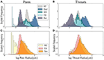
Effigy 5. Log normal distribution of the pore and throat geometry in sandstones (A,B) and limestones (C,D). The left column illustrate the distribution of pore radii, while the right column displays the distribution of throat radii. Six samples from this study (KF, Knorringfjellet; Wil, Wilcox; Dod, Doddington; MJ, Massangis Jaune; Sav, Savonnières; Est, Estaillades) and ii samples (FB, Fontainebleau; Ber, Berea) from Thomson et al. (2018).

Table 3. Pore network model characteristics for the macroporosity and permeability results.
The master attributes of the pore network display a power constabulary relationship with porosity. The data in these plots involve but the macroporosity. The plots shown in Effigy 6 demonstrate the human relationship between porosity and pore radius, throat radius and throat length in all 6 samples, with the addition of two sandstones used in a similar study past Thomson et al. (2018). We also plot the porosity-coordination number relation for comparison. In the log-log plots, we overlay fits of the form y = aϕ due north , in each plot with the value of northward annotated in the plots.
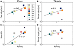
Figure 6. Macropore network characteristics of the six samples from this study (KF, Knorringfjellet; Wil, Wilcox; Dod, Doddington; MJ, Massangis Jaune; Sav, Savonnières; Est, Estaillades) and 2 samples (FB, Fontainebleau; Ber, Berea) from Thomson et al. (2018). The panels brandish log-log plots of (A) pore radius, (B) pharynx radius, (C) mean coordination number, and (D) pharynx length as a function of macroporosity. The broken line indicates a power constabulary fit through the six information points from this study. The ability law exponent for each fit is annotated in the plot. The ii samples with smaller symbol size are from college resolution images with smaller voxel dimensions.
Amid the PNM attributes considered here, the pore radius displays the strongest dependence on porosity. From the porosity-pore radius plot in Figure 6A we discover that the pore radius varies with porosity with an exponent of two/3. Both the radius and length of the throats in panels (B) and (D) evidence a well-nigh square-root variation with porosity. Coordination number of the pores besides increase with the porosity with an exponent of 0.57.
3.three. Absolute Permeability Simulation
We modeled the single phase flow of water directly through the pore space images of the macroporosity and total porosity cases forth the Z-axis just. Results of each simulation have been summarized in Table 3. In the Doddington sandstone, permeability measured through macroporosity has a value of 3,013 mD, which increases by 24% to 3,725 physician when the simulation is computed considering the total porosity. These are the highest values of permeability observed in the three sandstone samples. The Knorringfjellet sandstone shows a dramatic increase in permeability when considering the macroporosity and total porosity scenarios. The continued porosity proportion has a permeability of vii mD. The total porosity provided a value of 60 mD, which is 760% increment compared to the macroporosity only simulation. The Wilcox sandstone has zero connected porosity when analyzing the entire volume in this study, and therefore could not be used to simulate menstruation. Equally such, no permeability measurements were acquired. The Estaillades limestone allowed flow through its macroporosity at 259 physician. Compared to the total porosity model, this value increased by 132% to 601 mD. Similarly to the Wilcox sandstone, the Massangis Jaune limestone has zero connected porosity in the macroporosity case, meaning that no permeability simulation could exist modeled. However, in the total porosity case the connectivity across the Z-centrality permitted menstruum and a permeability measure out was obtained at 612 mD. The Savonnières limestone has the most efficient network for fluid flow, with a macroporosity permeability value at 1408 mD. The total porosity simulation provided an increased permeability at 3,990 mD, 183% more.
We display the combined porosity and permeability data in Figure vii. Where applicable, the plot displays data from both the macropore network (filled squares) and full pore network (filled circles). Also added in the plot are information from Fontainebleau and Berea sandstones from Thomson et al. (2018). We fit the data with an equation of the class . Values of both parameters are annotated in the plot. The exponent of northward = 3.32 from our data is higher than the theoretical value of n = 2 of throats with a circular cantankerous section, but is slightly lower than the value of three.8–iv.five obtained from experimental measurements (Doyen, 1988; Bernabé et al., 2010; Revil et al., 2014).
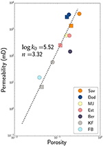
Figure 7. Plot of porosity and permeability from our samples and Fontainebleau (FB) and Berea (B) sandstones from Thomson et al. (2018). Filled circles stand for data from total porosity while filled squares represent data from macroporosity solitary. The broken line is a fit to the data of the form . The fit parameters are annotated in the plot.
4. Discussion
iv.1. Micropermeability
As discussed in the introduction, dimension of the micropores in our samples are across the limit of detection and assay in this study. As a result, certain assumptions must be fabricated regarding the geometry and connectivity of micropores Bultreys et al. (2015). Nosotros chose to select 2 end member cases. One time, the microporosity is considered impermeable, thus the permeability of the rock is determined only by the macroporosity. In the other case, nosotros consider the microporosity consists essentially of void space and the effective permeability of the stone is given past the total permeability. In reality, the microporous phase may have a non-zero permeability, rendering the effective permeability somewhere between these two cases. To rigorously address the issue of micropermeability ii problems need to be addressed in future work.
First, the 3D microtomographic images of the pore network can be supplemented by boosted, higher resolution 3D images, possibly obtained past techniques such as FIB electron microscopic tomography. These two dissimilar scales volition provide two values of permeability through the macro and micro pore spaces, which and so need to be averaged using some weighting formula.
Secondly, the nature of flow through nanometer-sized pores can be fundamentally dissimilar from the classic Darcy flow model. At such small pore dimensions, the ratio between area to volume of pores increment substantially (Israelachvili, 1991; Chen et al., 2004), rendering surface tension at the grain-fluid interface as a strong retarding strength to fluid percolation. Thus, evaluation of permeability from these pore spaces need to utilise a modified grade of Stokes flow, taking into account the surface tension (Hier-Majumder et al., 2006; Takei and Hier-Majumder, 2009; Hier-Majumder and Abbott, 2010).
Despite these uncertainties, the cease fellow member arroyo taken in this study tin be extremely useful in identifying the function of cementation in reducing permeability during reactive porous flow. In the following section, we discuss the implications of our results on such flows.
four.2. Upshot of Cementation on Fluid Transport
Cementation during reactive porous flow leads to the degradation of secondary phases along the initially interconnected pore network, reducing the constructive transport backdrop of the network. Using the results of our pore network models and permeability simulations, nosotros can quantify the influence of cement volume fraction on 2 important parameters, the fraction of connected pore infinite and the ratio between permeability afterward and before cementation.
The 3D image of Knoringfjellet sandstone in Figure 8 demonstrates the style cement, identified every bit the microporous phase, influences the pore space. The grain matrix is shown in gray scale, while the macroporosity (labeled "pore") and microporosity (labeled "cement") are identified in cyan and red, respectively. As the colored image on the right demonstrates, the cement fills up several channels essentially disconnecting the height and the bottom sections of the image. The presence of this cement has important consequences on the connectivity of the pore network.
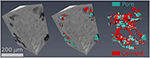
Figure 8. 3D visualization of gray calibration and segmented images of a subvolume in the Knorringfjellet sandstone. The micrograph in the centre overlaps the grey scale image on the segmented phases, whereas the micrograph on the right shows only the macropore and the microporous cement.
For instance, the Doddington sandstone has a total porosity of eighteen.6%, of which simply 1.ix% resides in microporosity (Tabular array 2). As a issue, 16.6% of the full volume of the Doddington sample resides in connected macropores. In contrast, Massangis Jaune carbonate shows a comparable total porosity of 16.one%, the majority of which (xi.2% of total volume) resides in microporosity in the cement. In this sample, no continued macroporosity was detected due to cementation. The plot in Effigy 9A plots the fraction of interconnected macropores as a role of cement volume fraction. As the plot depicts, the interconnectivity of the pore network is essentially reduced at cement book fractions greater than 4 vol%. This event implies that the connectivity of a pore network volition decline with fourth dimension as precipitation occurs during reactive porous menstruation, until this threshold value is reached at which bulk of the network will be cut-off from pervasive percolation of pore fluid.
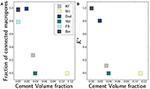
Figure nine. Plots of fraction of (A) interconnected macroporosity and (B) the ratio betwixt macro permeability and full permeability equally a role of cement volume fraction.
The outcome of cementation is likewise observed directly on the constructive permeability. Since the sectionalisation allows u.s.a. to evaluate the pore space before (full porosity) and after (macroporosity) cementation, the ratio of permeability between these two networks tin can be quite useful. The plot in Figure 9B illustrates this behavior in the plot of as a function of cement volume fraction. Every bit before, we notice that the permeability declines sharply with an increase in cement volume fraction, eventually becoming nix for cement volume fractions greater than 4 vol%. The permeability values are reported in Table three.
As permeability is progressively reduced by cement deposition, the fluid flux through a pore network will decrease with time. A number of recent studies on reactive porous menstruum during carbon capture and sequestration (Ghesmat et al., 2011; Unwin et al., 2016) and reaction infiltration instability (Takei and Hier-Majumder, 2009; Kelemen et al., 2011; Szymczak and Ladd, 2013) detailed the mode in which development of reaction fronts play an important role in sequestering dissolved chemical components in the fluid. None of these studies, however, accept into business relationship the way permeability changes during the progression of reactive porous menstruum. Every bit discussed above, the flux of porous period will diminish with time due to permeability reduction, thus reducing the efficiency of carbon sequestration over the life time of an performance. The reduction in flux of porous flow can also be seen as a positive, particularly in the upper regions of carbon sequestration reservoirs where the sealing capacity may increase with time due to cementation and clogging of pores. This tin can enhance reservoir storage and reduce unwanted leakage. Future models incorporating the influence of cementation volition provide more accurate and realistic estimates of the corporeality of carbon that can be stored in subsurface reservoirs.
5. Conclusions
• We analyzed 6 natural rocks, most containing a microporous cement stage. We consider the microporosity as void infinite to capture the pore network prior to cementation and solid to capture the pore geometry subsequently cementation.
• In the macropore space, the pore radius varies with porosity with an exponent of 2/3, while hateful coordination number, throat radius, and pharynx length all vary with a porosity exponent close to 1/2. Our results signal a porosity exponent of 3.3 for permeability.
• We find that, within the sample set up used in this study and the resolution of our images, the fraction of connected pores and permeability subtract sharply with cement volume fraction and the connectivity of macropores is cut off virtually a cement volume fraction of 4%.
Data Availability
The results from PNM model are available through Figshare: Hier-Majumder, Saswata; Thomson, Paul-Ross; Hazel, Alexander (2019): Information for "The Influence of Microporous Cements on the Pore Network Geometry of Natural Sedimentary Rocks." figshare. Fileset. https://doi.org/10.17637/rh.7667642.
Author Contributions
P-RT carried out the image segmentation and permeability simulations in all samples. AH carried out the analysis and permeability simulations on Doddington and Knorringfjellet sandstones. SH-M and P-RT jointly carried out the final pore-network modeling and statistical assay. All 3 authors contributed to writing and revision of the manuscript.
Funding
This research was as well supported by the grant EAR125880 from the United states of america National Scientific discipline Foundation.
Conflict of Interest Argument
The authors declare that the research was conducted in the absence of any commercial or financial relationships that could exist construed equally a potential conflict of involvement.
Acknowledgments
The authors acknowledge insightful suggestions from Yiqiao Wu on the pore network models of the carbonate rocks. The raw 3D Images used in this work were kindly provided past Tom Bultreys. P-RT acknowledges support from a NERC Oil and Gas CDT graduate fellowship (grant number NE/M00578X/1).
Supplementary Cloth
The Supplementary Fabric for this article can be found online at: https://www.frontiersin.org/articles/10.3389/feart.2019.00048/full#supplementary-fabric
References
Alyafei, N., and Edgeless, M. J. (2016). The consequence of wettability on capillary trapping in carbonates. Adv. H2o Resour. 90, 36–l. doi: 10.1016/j.advwatres.2016.02.001
CrossRef Full Text | Google Scholar
Andrä, H., Combaret, Due north., Dvorkin, J., Glatt, E., Han, J., Kabel, M., et al. (2013a). Digital rock physics benchmarks-Function I: imaging and segmentation. Comput. Geosci. 50, 25–32. doi: ten.1016/j.cageo.2012.09.005
CrossRef Full Text | Google Scholar
Andrä, H., Combaret, Due north., Dvorkin, J., Glatt, E., Han, J., Kabel, 1000., et al. (2013b). Digital rock physics benchmarks-part II: Calculating effective properties. Comput. Geosci. l, 33–43. doi: 10.1016/j.cageo.2012.09.008
CrossRef Total Text | Google Scholar
Bera, B., Mitra, S. Chiliad., and Vick, D. (2011). Agreement the micro structure of Berea Sandstone by the simultaneous use of micro-computed tomography (micro-CT) and focused ion beam-scanning electron microscopy (FIB-SEM). Micron 42, 412–418. doi: 10.1016/j.micron.2010.12.002
PubMed Abstract | CrossRef Full Text | Google Scholar
Berg, C. F., Lopez, O., and Berland, H. (2017). Industrial applications of digital rock engineering. J. Petrol. Sci. Eng. 157, 131–147. doi: 10.1016/j.petrol.2017.06.074
CrossRef Full Text | Google Scholar
Bernabé, Y., Li, One thousand., and Maineult, A. (2010). Permeability and pore connectivity: a new model based on network simulations. J. Geophys. Res. Solid World 115, 1–14. doi: 10.1029/2010JB007444
CrossRef Full Text
Boone, Thousand. A., De Kock, T., Bultreys, T., De Schutter, G., Vontobel, P., Van Hoorebeke, L., et al. (2014). 3D mapping of h2o in oolithic limestone at atmospheric and vacuum saturation using 10-ray micro-CT differential imaging. Mater. Grapheme. 97, 150–160. doi: 10.1016/j.matchar.2014.09.010
CrossRef Total Text | Google Scholar
Bultreys, T., De Boever, Due west., and Cnudde, V. (2016a). Imaging and image-based fluid ship modeling at the pore scale in geological materials: a practical introduction to the electric current state-of-the-art. Earth Sci. Rev. 155, 93–128. doi: x.1016/j.earscirev.2016.02.001
CrossRef Total Text | Google Scholar
Bultreys, T., Stappen, J. V., Kock, T. D., Boever, W. D., Boone, Yard. A., Hoorebeke, Fifty. Five., et al. (2016b). Investigating the relative permeability beliefs of microporosity-rich carbonates and tight sandstones with multiscale pore network models. J. Geophys. Res. Solid Earth 121, 7929–7945. doi: 10.1002/2016JB013328
CrossRef Total Text | Google Scholar
Bultreys, T., Van Hoorebeke, L., and Cnudde, V. (2015). Multi-scale, micro-computed tomography-based pore network models to simulate drainage in heterogeneous rocks. Adv. Water Resour. 78, 36–49. doi: 10.1016/j.advwatres.2015.02.003
CrossRef Total Text | Google Scholar
Chen, Northward., Kuhl, T., Tadmor, R., Lin, Q., and Israelachvili, J. (2004). Big deformation during coalescence of fluid interfaces. Phys. Rev. Lett. 92:024501. doi: 10.1103/PhysRevLett.92.024501
CrossRef Full Text | Google Scholar
Choquette, P. W., and Pray, L. C. (1970). Geologic classification and classification of porosity in sedimentary carbonates. AAPG Bull. 54, 207–250.
Google Scholar
Cnudde, V., and Boone, Grand. N. (2013). Loftier-resolution 10-ray computed tomography in geosciences: a review of the electric current applied science and applications. Earth Sci. Rev. 123, 1–17. doi: 10.1016/j.earscirev.2013.04.003
CrossRef Full Text | Google Scholar
Derluyn, H., Dewanckele, J., Boone, M. Due north., Cnudde, V., Derome, D., and Carmeliet, J. (2014). Crystallization of hydrated and anhydrous salts in porous limestone resolved past synchrotron X-ray microtomography. Nuclear Instrum. Methods Phys. Res. B 324, 102–112. doi: ten.1016/j.nimb.2013.08.065
CrossRef Full Text | Google Scholar
Doyen, P. M. (1988). Permeability, conductivity, and pore geometry of sandstone. J. Geophys. Res. 93:7729.
Google Scholar
Ghesmat, K., Hassanzadeh, H., and Abedi, J. (2011). The impact of geochemistry on convective mixing in a gravitationally unstable diffusive boundary layer in porous media: CO2storage in saline aquifers. J. Fluid Mech. 673, 480–512. doi: 10.1017/S0022112010006282
CrossRef Full Text | Google Scholar
Hier-Majumder, Due south., and Abbott, M. E. (2010). Influence of dihedral bending on the seismic velocities in partially molten rocks. World Planet. Sci. Lett. 299, 23–32. doi: x.1016/j.epsl.2010.08.007
CrossRef Total Text | Google Scholar
Hier-Majumder, S., Ricard, Y., and Bercovici, D. Role of grain boundaries in magma migration storage. (2006). Earth Planet. Sci. Lett. 248, 735–749. doi: 10.1016/j.epsl.2006.06.015
CrossRef Full Text
Iassonov, P., Gebrenegus, T., and Tuller, M. (2009). Segmentation of x-ray computed tomography images of porous materials: a crucial step for label and quantitative analysis of pore structures. Water Resour. Res. 45:W09415. doi: 10.1029/2009WR008087
CrossRef Full Text | Google Scholar
Israelachvili, J. Due north. (1991). Intermolecular and Surface Forces, 2 Edn. London; San Diego, CA: Academic Printing.
Jennings, J. Due west., and Lucia, F. J. (2003). Predicting Permeability From Well Logs in Carbonates With a Link to Geology for Interwell Permeability Mapping. SPE Reservoir Evaluation & Applied science. Vol. 6. Society of Petroleum Engineers. doi: x.2118/84942-PA
CrossRef Full Text
Jiang, Z., Van Dijke, Yard. I., Sorbie, Yard. S., and Couples, K. D. (2013). Representation of multiscale heterogeneity via multiscale pore networks. Water Resour. Res. 49, 5437–5449. doi: ten.1002/wrcr.20304
CrossRef Full Text | Google Scholar
Jiang, Z., van Dijke, M. I., Wu, G., Couples, One thousand. D., Sorbie, K. S., and Ma, J. (2012). Stochastic pore network generation from 3D rock images. Transport Porous Media 94, 571–593. doi: 10.1007/s11242-011-9792-z
CrossRef Full Text | Google Scholar
Jiao, M., Yao, S., Liu, C., Gao, Y., Wu, H., Li, M., and Tang, Z. (2014). The label and quantitative analysis of nanopores in unconventional gas reservoirs utilizing FESEM-FIB and prototype processing: an example from the lower Silurian Longmaxi Shale, upper Yangtze region, Mainland china. Int. J. Coal Geol. 128-129, i–11. doi: ten.1016/j.coal.2014.03.004
CrossRef Full Text | Google Scholar
Kelemen, P. B., Matter, J., Streit, E. E., Rudge, J. F., Curry, W. B., and Blusztajn, J. (2011). Rates and mechanisms of mineral carbonation in peridotite: natural processes and recipes for enhanced, in situ CO2 capture and storage. Annu. Rev. Earth Planet. Sci 39 545–576. doi: 10.1146/annurev-earth-092010-152509
CrossRef Full Text | Google Scholar
Ketcham, R. A., and Carlson, Due west. D. (2001). Acquisition, optimization and interpretation of x-ray computed tomographic imagery: applications to the geosciences. Comput. Geosci. 27, 381–400. doi: 10.1016/S0098-3004(00)00116-3
CrossRef Full Text | Google Scholar
Ma, L., Taylor, K. G., Lee, P. D., Dobson, K. J., Dowey, P. J., and Courtois, Fifty. (2016). Novel 3D centimetre-to nano-scale quantification of an organic-rich mudstone: The Carboniferous Bowland Shale, Northern England. Mar. Petrol. Geol. 72, 193–205. doi: 10.1016/j.marpetgeo.2016.02.008
CrossRef Full Text | Google Scholar
Madonna, C., Quintal, B., Frehner, M., Almqvist, B. Southward. G., Tisato, Due north., Pistone, 1000., et al. (2013). Synchrotron-based 10-ray tomographic microscopy for rock physics investigations. Geophysics 78, D53–D64. doi: 10.1190/geo2012-0113.1
CrossRef Full Text | Google Scholar
Marquez, X., Solling, T., Finlay, Due south., Bounoua, Northward., and Gagigi, T. (2014). 3D imaging of porosity modifying phases in Shuaiba Reservoir, Al Shaheen Field. International Petroleum Applied science Conference (Doha), sixteen.
Google Scholar
Mehmani, A., and Prodanović, Grand. (2014). The effect of microporosity on transport properties in porous media. Adv. Water Resour. 63, 104–119. doi: x.1016/j.advwatres.2013.10.009
CrossRef Full Text | Google Scholar
Menke, H., Andrew, 1000., Blunt, M., and Bijeljic, B. (2016). Reservoir condition imaging of reactive transport in heterogeneous carbonates using fast synchrotron tomography—effect of initial pore construction and menstruum conditions. Chem. Geol. 428, 15–26. doi: ten.1016/j.chemgeo.2016.02.030
CrossRef Full Text | Google Scholar
Menke, H. P., Bijeljic, B., Andrew, 1000. Yard., and Blunt, M. J. (2015). Dynamic three-dimensional pore-scale imaging of reaction in a carbonate at reservoir conditions. Environ. Sci. Technol. 49, 4407–4414. doi: ten.1021/es505789f
PubMed Abstract | CrossRef Total Text | Google Scholar
Mostaghimi, P., Blunt, M. J., and Bijeljic, B. (2013). Computations of accented permeability on micro-CT images. Math. Geosci. 45, 103–125.
Google Scholar
Okabe, H., and Blunt, Chiliad. J. (2005). Pore space reconstruction using multiple-point statistics. J. Petrol. Sci. Eng. 46, 121–137. doi: x.1007/s11004-012-9431-four
CrossRef Full Text | Google Scholar
Øren, P.-E., and Bakke, S. (2003). Reconstruction of berea sandstone and pore-scale modelling of wettability furnishings. J. Petrol. Sci. Eng. 39, 177–199. doi: 10.1016/j.petrol.2004.08.002
CrossRef Total Text | Google Scholar
Pal, N. R., and Pal, Southward. K. (1993). A review on image segmentation techniques. Pattern Recogn. 26, 1277–1294. doi: 10.1016/S0920-4105(03)00062-7
CrossRef Full Text | Google Scholar
Pittman, Due east. D. (1979). "Porosity, diagenesis and productive adequacy of sandstone reservoirs," in 'Aspects of Diagenesis', (SEPM Society for Sedimentary Geology). doi: 10.2110/pec.79.26.0159
CrossRef Full Text | Google Scholar
Prodanović, Thou., Mehmani, A., and Sheppard, A. P. (2014). Imaged-based multiscale network modelling of microporosity in carbonates. Geol. Soc. Lond. 406, SP406–SP409. doi: x.1016/0031-3203(93)90135-J
CrossRef Total Text
Raeini, A. Q., Blunt, M. J., and Bijeljic, B. (2014). Direct simulations of two-phase catamenia on micro-CT images of porous media and upscaling of pore-scale forces. Adv. Water Resour. 74, 116–126. doi: ten.1016/j.advwatres.2014.08.012
CrossRef Full Text | Google Scholar
Ramstad, T., Øren, P.-E., Bakke, S., et al. (2010). Simulation of two-phase catamenia in reservoir rocks using a lattice boltzmann method. SPE J. fifteen, 917–927. doi: 10.2118/124617-PA
CrossRef Full Text | Google Scholar
Revil, A., Kessouri, P., and Torres-Verdín, C. (2014). Electrical electrical conductivity, induced polarization, and permeability of the Fontainebleau sandstone. Geophysics 79, D301–D318. doi: 10.1190/geo2014-0036.one
CrossRef Full Text | Google Scholar
Saif, T., Lin, Q., Butcher, A. R., Bijeljic, B., and Blunt, K. J. (2017). Multi-scale multi-dimensional microstructure imaging of oil shale pyrolysis using X-ray micro-tomography, automated ultra-loftier resolution SEM, MAPS Mineralogy and FIB-SEM. Appl. Ener. 202, 628–647. doi: 10.1016/j.apenergy.2017.05.039
CrossRef Total Text | Google Scholar
Schlüter, S., Sheppard, A., Brown, K., and Wildenschild, D. (2014). Image processing of multiphase images obtained via Ten-ray microtomography: a review. Water Resour. Res. 50, 3615–3639. doi: 10.1002/2014WR015256
CrossRef Full Text | Google Scholar
Sezgin, Grand., and Sankur, B. (2004). Survey over prototype thresholding techniques and quantitative functioning evaluation. J. Electron. Imaging 13, 146–166. doi: x.1117/i.1631315
CrossRef Full Text | Google Scholar
Shah, S., Greyness, F., Crawshaw, J., and Boek, E. (2016). Micro-computed tomography pore-scale study of catamenia in porous media: Effect of voxel resolution. Adv. H2o Resour. 95, 276–287. doi: 10.1016/j.advwatres.2015.07.012
CrossRef Full Text | Google Scholar
Szymczak, P., and Ladd, A. J. C. (2013). Interacting length scales in the reactive-infiltration instability. Geophys. Res. Lett. 40, 3036–3041. doi: ten.1002/grl.50564
CrossRef Full Text | Google Scholar
Takei, Y., and Hier-Majumder, S. (2009). A generalized formulation of interfacial tension driven fluid migration with dissolution/precipitation. Globe Planet. Sci. Lett. 288, 138–148. doi: 10.1016/j.epsl.2009.09.016
CrossRef Full Text | Google Scholar
Thomson, P.-r., Aituar-zhakupova, A., and Hier-majumder, S. (2018). Image segmentation and analysis of pore network geometry in two natural sandstones. Front. Earth Sci. half-dozen:58. doi: 10.3389/feart.2018.00058
CrossRef Full Text | Google Scholar
Trier, Ø. D., and Jain, A. 1000. (1995). Goal-directed evaluation of binarization methods. IEEE Trans. Patt. Anal. Mach. Intelligence, 12, 1191–1201.
Google Scholar
Unwin, H. J. T., Wells, Grand. Northward., and Wood, A. W. (2016). CO2 dissolution in a background hydrological menstruation. J. Fluid Mech. 789, 768–784. doi: 10.1017/jfm.2015.752
CrossRef Full Text | Google Scholar
Valvatne, P. H., and Blunt, G. J. (2004). Predictive pore-scale modeling of 2-stage menses in mixed wet media. H2o Resour. Res. 40:W07406. doi: ten.1029/2003WR002627
CrossRef Total Text | Google Scholar
Wang, W., Kravchenko, A., Smucker, A., and Rivers, One thousand. (2011). Comparison of image segmentation methods in false 2d and 3d microtomographic images of soil aggregates. Geoderma 162, 231–241. doi: 10.1016/j.geoderma.2011.01.006
CrossRef Full Text | Google Scholar
Wildenschild, D., and Sheppard, A. P. (2013). X-ray imaging and analysis techniques for quantifying pore-calibration construction and processes in subsurface porous medium systems. Adv. Water Resour. 51, 217–246. doi: 10.1016/j.advwatres.2012.07.018
CrossRef Full Text | Google Scholar
Wildenschild, D., Vaz, C., Rivers, 1000., Rikard, D., and Christensen, B. (2002). Using x-ray computed tomography in hydrology: systems, resolutions, and limitations. J. Hydrol. 267, 285–297. doi: 10.1016/S0022-1694(02)00157-9
CrossRef Total Text | Google Scholar
Youssef, South., Rosenberg, E., Gland, N. F., Kenter, J. A., Skalinski, One thousand., and Vizika, O. (2007). "High resolution CT and pore-network models to assess petrophysical properties of homogeneous and heterogeneous carbonates," in SPE/EAGE Reservoir Characterization and Simulation Briefing, 28–31 Oct (Abu Dhabi: Society of Petroleum Engineers), 28-31. doi: 10.2118/111427-MS
CrossRef Total Text | Google Scholar
Source: https://www.frontiersin.org/articles/10.3389/feart.2019.00048/full
0 Response to "A Review of the Nature and Origin of Limestone Microporosity"
Post a Comment